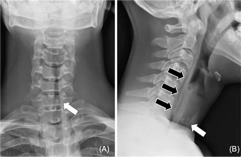
normal soft tissue neck x-ray|soft tissue neck x ray positioning : Pilipinas Normal soft tissue neck x-ray: lateral projection. Case contributed by Henry . The Bachelorette season 20 star Charity Lawson gives a post-finale update with Dotun Olubeko after falling in love with new Bachelor Joey Graziadei.
PH0 · soft tissue x ray technique
PH1 · soft tissue neck x ray protocol
PH2 · soft tissue neck x ray positioning
PH3 · soft tissue neck x ray kvp
PH4 · soft tissue neck x ray anatomy
PH5 · soft tissue neck anatomy
PH6 · lateral soft tissue neck x ray
PH7 · ap soft tissue neck x ray
PH8 · Iba pa
We would like to show you a description here but the site won’t allow us.
normal soft tissue neck x-ray*******Soft tissue neck. x-ray. Normal delineation of the pharynx, larynx, and trachea. Paravertebral soft tissues demonstrate normal width, no evidence of foreign bodies or gas in the soft tissues. Cervical spine vertebral bodies have normal height and alignment.Lateral soft tissue radiograph of the neck demonstrates thickening of the epiglottis .
Similarly normal values for CT vary, but according to one of the larger series in .
Normal soft tissue neck x-ray: lateral projection. Case contributed by Henry .
Normal soft-tissue neck x-ray. A soft-tissue neck series consists of an anterior–posterior (AP) (A) and a lateral (B) x-ray of the neck. Compared with a . Radiograph showing the soft tissues of the neck: lateral view. Name the areas labelled A-F that are important when looking at a .Soft Tissue Neck X-ray Guideline. Soft Tissue Neck: 2views • AP and LATERAL • AP neck. 40” and 15° cephalic angle. Take film during phonation or crying (infants). • .
normal soft tissue neck x-ray Soft Tissue Neck Radiographs. March 15, 2015. The soft-tissue neck radiograph can be an extremely useful tool in a variety of clinical situations. These include: epiglottitis, croup, retropharyngeal .
10.1055/b-0036-138090 17 Neck Soft Tissue 17.1 Introduction The soft tissues of the neck can be an intimidating area to evaluate radiologically. However, children have numerous congenital .patient appropriately. A lateral soft tissue neck radiograph is a cheap, readily available investigation tool that is of clinical value in assessing patients with potential pathology of . A normal neck X-ray should display certain key features: • Alignment: The cervical vertebrae should align correctly, forming a gentle, natural curve. • Bone .
Soft Tissue Neck X-ray Guideline. Soft Tissue Neck: 2views • AP and LATERAL • AP neck. 40” and 15° cephalic angle. Take film during phonation or crying (infants). • LATERAL neck. 72” Extend neck. Take film during inspiration with mouth closed. • If evaluation of adenoids is requested do lateral sinus on inspiration with mouth closed
UQ anatomy presentation - Normal Anatomy by Lachlan Inch; Normal Anatomy by Stallon Sebastian; RACS/UQ Advanced Surgical Anatomy Course - Brain, neck, spine by Craig Hacking ANZCA workshop 2017 by Craig Hacking C-Spine by Bryan Schnabel Normal rx by Sabrina Martel; Spine by Luke Abela; NRA Spine and pelvis by Tom Molyneux Gross anatomy. The perivertebral space is a cylinder of soft tissue lying posterior to the retropharyngeal space and danger space surrounded by the prevertebral layer of the deep cervical fascia and .An AP soft tissue of neck x-ray is an important diagnostic tool. It can reveal tumors, masses, or swallowed foreign objects. This image also shows the position of the esophagus and upper airway. If you have any doubts about the results of your neck x-ray, talk to your doctor. This image has many advantages over x-ray.
The thickness of the prevertebral soft tissue (PVST) has long been considered a valuable radiographic measurement in evaluating possible injury to the cervical spine. 1–6 Analysis of the PVST is helpful in detecting subtle osseous or ligamentous injuries that might go unrecognized. In our experience, the normal values .An x-ray soft-tissue neck positioning sequence consists of two types of images: the anterior-posterior (AP) and lateral (B) views of the neck. These images . Abnormal soft-tissue x-rays may reveal air in the neck, deviation from normal air-filled structures, or compression of bones. X-rays may also detect radiopaque objects, although they are .soft tissue neck x ray positioningAge: 35 years. Gender: Female. x-ray. Normal examination. Prevertebral soft tissues are within normal limits. No radiopaque foreign body.

Role of plain xray soft tissue neck lateral view in the diagnosis of cervical esophageal foreign bodies. Internet J Otorhinolaryngol 2008;8(2):1–5. Google Scholar; 41. Anderson KL, Dean AJ. Foreign bodies in the gastrointestinal tract and anorectal emergencies. Emerg Med Clin North Am 2011;29(2):369–400, ix. Crossref, Medline, . posterior nasal space x-ray: example needed. soft tissue neck. 6-year-old: example 1. CT. CT cervical spine: 3-year-old: example 1. 7-year-old: example 1. 12-year-old: example 1. 13-year-old: example 1. CT neck: example needed. MRI. MRI neck: example needed. Thoracolumbar spine Plain radiograph. thoracic spine. 6-year-old: . These structures will appear dark gray on the X-ray image. Soft tissues include: . where the muscles and ligaments of the neck are forced to move outside their normal range. If your neck is .Neck X-rays are a form of radiographic imaging that focuses on the cervical spine and the surrounding structures of the neck. They are widely used to evaluate bone and soft tissue structures in the neck region. These X-rays are instrumental in diagnosing and monitoring various medical conditions related to the neck and upper spine.
When interpreting an X-ray of soft-tissue neck, never forget to comment on these points: a. Cervical vertebrae: Erosion of vertebral bodies; Loss of cervical lordosis (due to prevertebral muscle spasm) b. .
First, soft-tissue neck x-rays are generally underexposed. This is done to better examine the soft tissue. Specifically, a soft-tissue neck x-ray will show a thin strip of air in the retropharyngeal space, which extends from .The thickness of the prevertebral soft tissue (PVST) has long been considered a valuable radiographic measurement in evaluating possible injury to the cervical spine. 1–6 Analysis of the PVST is helpful in detecting subtle osseous or ligamentous injuries that might go unrecognized. In our experience, the normal values based on radiographic studies are .In addition, he shows air-fluid level in the retropharyngeal space. A comparison of the x-ray with a normal soft-tissue neck image is provided in the following figure. Epiglottitis. The diagnosis of epiglottitis after soft tissue neck X-ray has traditionally been made on qualitative radiographic signs. However, a recent study evaluated the .

Normal soft tissue neck radiograph Case contributed by Craig Hacking. Diagnosis not applicable Share Add to Citation, DOI, disclosures and case data. Citation: . Soft tissue Neck X-Ray by Ahmed Mohamed Mohamed Eid Ali; Related Radiopaedia articles. Normal head and neck imaging examples; Promoted articles (advertising)Age: 15 years. Gender: Male. x-ray. Normal lateral neck radiograph. The hyoid is visible, but no calcification of the neck cartilage structures is present yet. No prevertebral soft tissue thickening. Normal cervical spine.normal soft tissue neck x-ray soft tissue neck x ray positioningAge: 35 years. Gender: Female. x-ray. Normal examination. Prevertebral soft tissues are within normal limits. No radiopaque foreign body.
New art movies to stream, including documentaries on M.C. Escher, Marcel Duchamp, Hilma af Klint, David Wojnarowicz, Banksy, and Pat Steir.
normal soft tissue neck x-ray|soft tissue neck x ray positioning